
Skeleton
A model skeleton accurately scaled in terms of height and proportions, with approximately 200 numbered bones, a flexible vertebral column, and a removable three-part skull. It is on a rolling stand, and can be easily moved to small group study areas or other study spaces.
H eart
eart
Approximately two times natural size. Sectioned so that both ventricles and atria open to expose the valves. Large blood vessels near the heart and musculature of the heart are shown. Separates into 4 parts.
 Brain with arteries
Model of brain with representation of arterial network of vessels. Natural size. Separates into 9 parts: frontal and parietal lobes (2), temporal and occipital lobes (2), medulla (2), cerebellum (2), and basilar artery.
 Pelvis model 1
Female genital and pelvic organs with bladder and rectum fully exposed and removable. Natural size. Separates into 2 parts.
 Pelvis model 2
Male pelvic and genital organs with rectum, bladder with prostate and testicular duct and external genital organs, fully exposed, and removable. Natural size. Separates into 4 parts.
 Brain stem

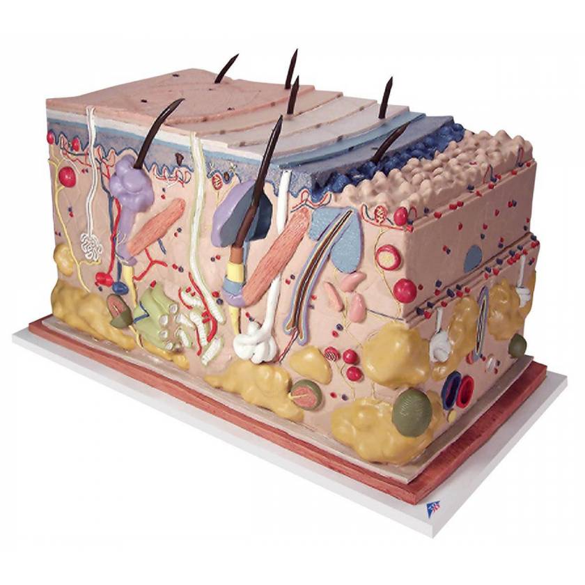
This model shows a section of human skin in three dimensional form. ndividual skin layers are differentiated, and important structures such as hair, sebaceous and sweat glands, receptors, nerves and vessels are shown in detail. Mounted on baseboard. Has accompanying key.

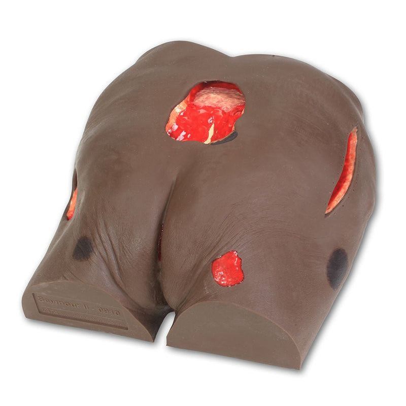
Wound care model
Molded from a 74-year-old patient and displaying the following conditions: Stage I, Stage II, Stage III with undermining, tunneling, subcutaneous fat and slough, deep Stage IV with exposed bone, undermining, tunneling, subcutaneous fat, eschar and slough. Also shown are a suspected DTI, unstagable, and a 5 1/2" dehisced wound. The unique flexible life-like material permits the placement of dressings for use with negative pressure wound therapy devices without leaving an adhesive residue. Wound cleansing, classification and staging as well as the measurement of wounds can be demonstrated.